Virtual microscopy or digital microscopy is the digital conversion of light microscopic specimens in full resolution and their presentation over a computer network. In most cases the specimens are scanned with special microscopes, so-called slide scanners, but they can also be produced by joining several photos taken through the microscope.
The frequently used term “virtual” refers to the examination of the specimens without direct contact to the object slide or the light microscope.
The term “high-resolution digital microscopy” is more precise, but not frequently used. Other terms that are frequently being used are: internet based microscopy, ePathology, eHistology, eHematology, eBiology or virtual microscope. These terms are to some extent used synonymously with the term virtual microscopy.
Microscope slides are digitalized either manually or fully automatically with a 20x or 40x objective and saved in various formats, depending on the different service providers. All formats allow for relative fast visualisation of the specimens through a software specifically designed for this application (the so-called client or viewer) that is locally installed. The size of the image files that are created by scanning can vary between 5 megabytes (MB) and several gigabytes (GB), depending on the size of the specimen. The images for virtual microscopy can be saved on the local device. But it is more common to store them on a server with a large storage capacity so that the images can be accessed via an internet connection. For this purpose special software solutions were developed that allow a visualisation with a common web browser. (see section “Technology”). These HTML-based solutions make the installation of special software on the local device unnecessary and are usually barrier-free, meaning computer and operating system independent. For an optimal speed of image transmission a well thought through segmentation and an extremely optimized management of the large image files and their levels of magnification into smaller image tiles is necessary, so that the images and their zoom levels are called up selectively and only on demand of the user. Virtual microscopy could therefore be considered a sort of “streaming on demand” of image information in different zoom levels and at different topographical regions of the original image. The original file is at no time completely downloaded or locally stored.
The rate of data transmission rate depends on:
- the size of the smallest image subunit or “image tile”,
- the software used for image editing or rather for the segmentation into smallest picture subunits,
- the speed of the network and
- the performance of the terminal device
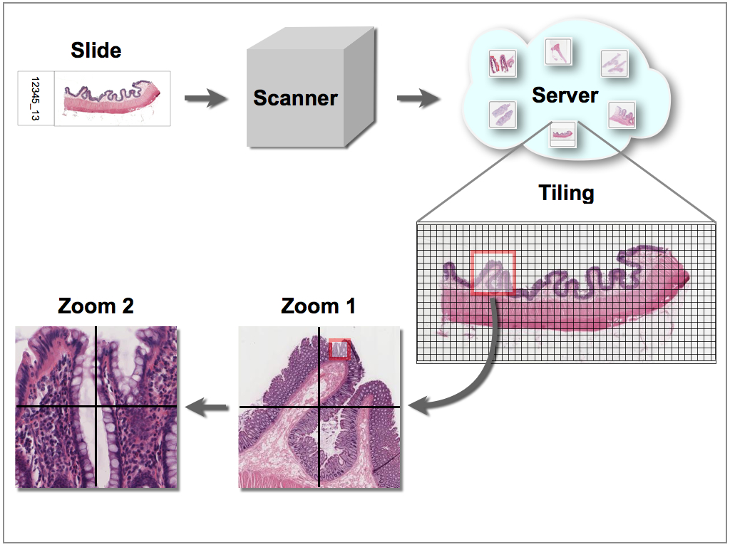
Figure 1: Schematic depiction of the digitalization of a histological specimen with a slide scanner for virtual microscopy. The original image is segmented into smaller subunits that can be accessed at different zoom levels via the internet or an internal computer network. For example, an 1 GB image can be segmented into smaller images (as small as 10 KB), so that only regions of the specimen with the according zoom level need to be called up by the user.